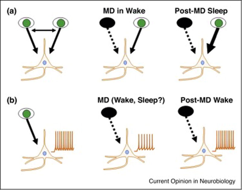
Figure 2. The effects of sleep on ODP in binocular cortex and homeostatic plasticity in monocular cortex are different.

In the binocular visual cortex of cats, MD of the ipsilateral or contralateral eye in the awake animal initially causes a breakdown of binocular responses and a weakening of circuits serving the deprived eye. During subsequent sleep, the response to the non-deprived eye is strengthened via mechanisms similar to Hebbian LTP (A). In the monocular visual cortex of rats, MD of the contralateral eye causes a drop in spontaneous activity, as the contralateral eye provides the sole input to monocular cortex. This is best explained by Hebbian long-term depression. The role of sleep and wake in this initial plastic change is currently unknown. Spontaneous activity slowly recovers (‘upscales’) over the next 2 days. This latter form of homeostatic plasticity is inhibited by sleep (B).
