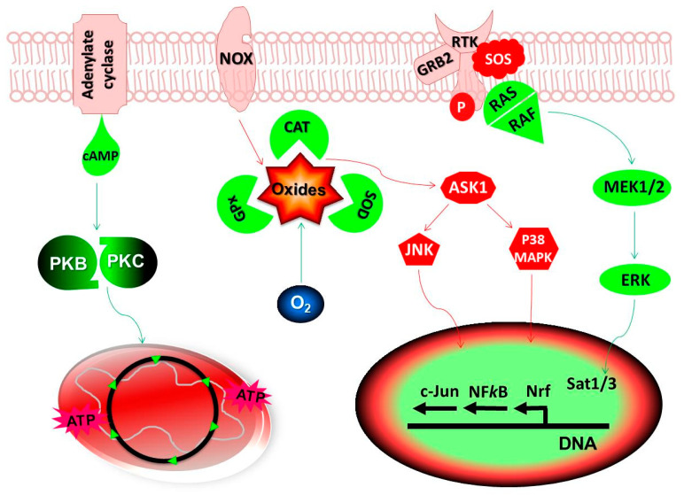
Figure 1.
Redox potential of antioxidants: In healthy cells, oxide free radicals generated from mitochondria and cell membrane NADPH Oxidases (NOX) are detoxified by the endogenous antioxidants, catalases (CAT), Glutathione Peroxidases (GPx) and Superoxide Dismutases (SOD). A constitutively low level of Reactive Oxygen Species (ROS) stimulate Protein Kinases (PK) B/C for maintaining cellular homeostasis while, higher levels can result in the change in mitochondrial membrane potential leading to apoptosis or cell death. Cancer cells have higher levels of oxidative stress as compared to the normal cells allowing them to activate several signaling pathways and transcription factors. Any imbalances in the redox-sensitive signaling pathways, Mitogen Activated Protein Kinases (MAPK) and PK B/C may result in the tumor growth. The Ras-Raf-MEK-ERK pathway (also known as the MAPK/ERK pathway) is the signaling cascade from cell membrane receptor complex to the nucleus. During excessive oxidative stress, Apoptosis Signal-regulating Kinase 1 (Ask1) induces apoptosis by activating c-Jun N-terminal Kinases (JNK) and p38 MAPK pathways. It may also lead to the inactivation and proteolytic degradation of PK molecules leading to the initiation of apoptosis or cell death signaling. The redox regulation of cellular oxidative stress is achieved either by activation or repression of the antioxidant genes through several transcription factors, acting individually or in combination. Objects in RED show the pathways activated directly by oxides radicals and the objects in GREEN indicate pathways regulating the cellular antioxidant mechanisms. Abbreviations: cAMP, cyclic Adenosine Monophosphate; ATP, Adenosine Triphosphate; ERK, Extracellular Signal-regulated Kinases; O2, oxygen.

