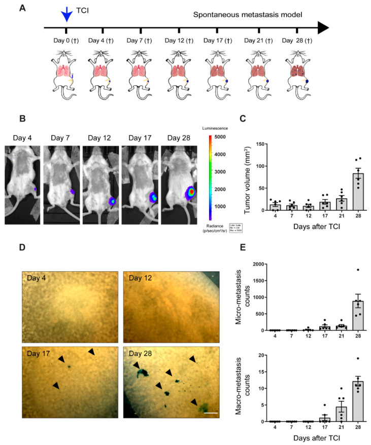
Figure 1.
Characterization of spontaneous metastasis in an orthotopic 143-B osteosarcoma model. (A) Scheme of the experimental setup, with abbreviations: tumor cell injection (TCI); euthanasia (†). (B) Representative IVIS bioluminescence images of tumor-bearing animals sacrificed at the indicated time points. (C) Primary tumor growth of 143-B cells over time, monitored by caliper measurements of the tumor volume at indicated time points. (D) Representative images of X-gal stained metastases on lung surface of mice sacrificed at indicated time points after TCI. Scale bar, 500 µm. (E) Quantification of pulmonary micro-metastasis (<0.1 mm in diameter, top panel,) and macro-metastasis (>0.1 mm in diameter, bottom panel) in mice sacrificed at indicated time points. n = 6 per time point.

