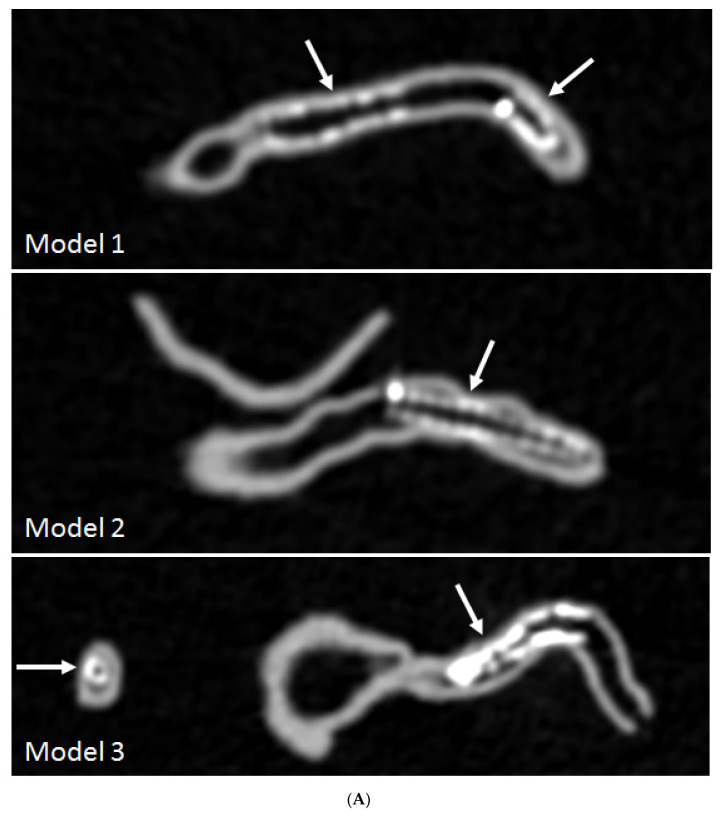
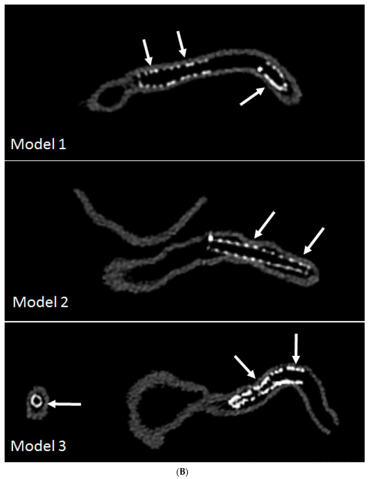
Figure 7.
3D printed coronary models with coronary stenting scanned on 192-slice CT scanner and images reconstructed with soft and sharp kernel algorithms. (A) images were reconstructed with standard soft kernel Bv36 algorithm showing that right coronary stents in these 3 models and left anterior descending stent (in model 3). (B) images were reconstructed with sharp kernel Bv59 of the same models with significant improvement of visualization of the stents and stented lumen, in particular improved visualization of stent wires in models 2 and 3. Arrows refer to the stent wires. Reprinted with permission under open access from Sun and Jansen [63].]


