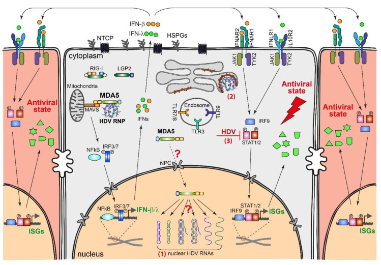
Figure 4.
HDV-induced IFN response and possible HDV countermeasures. HDV RNA in the cytoplasm; likely, the RNP complex is recognized by the pattern recognition receptor (PRR) MDA5 [106]. This recognition activates the mitochondrial antiviral signaling protein (MAVS) on the mitochondria and downstream transcription factors, likely IFN regulatory factor (IRF) 3/7 and nuclear factor-κB (NFκB). The activated transcription factors are translocated into the nucleus and initiate the transcription of IFN-β/λ. Secreted IFN-β/λ binds to their receptors (IFNAR1/IFNAR2 for IFN-α/β and IFNLR1/IL10R2 for IFN-λ) on the infected cell or neighboring cells, which further activates Janus kinases (JAK) 1/2, tyrosine kinase (TYK) 2, and transcription factors signal transducer and activator of transcription (STAT) 1/2 and IRF9. STAT1/2 and IRF9 are translocated into the nucleus and activate hundreds of IFN-stimulated genes (ISGs), which directly inhibit HDV replication and protect the uninfected cells against subsequent infection. It is unknown whether MDA5 can also be transported to the nucleus and capture nuclear HDV replication intermediates. HDV may counteract the IFN response through different strategies: (1) HDV may replicate in a “safe” compartment, the nucleus, to avoid exposure of the replication intermediates to PRRs; (2) HDV genomic RNA in the cytoplasm may fold into RNP with HDAg and bud into an HBV envelope to avoid being recognized by the PRRs; and (3) HDV may directly inhibit STAT1/2 activation.

