Figure 1.
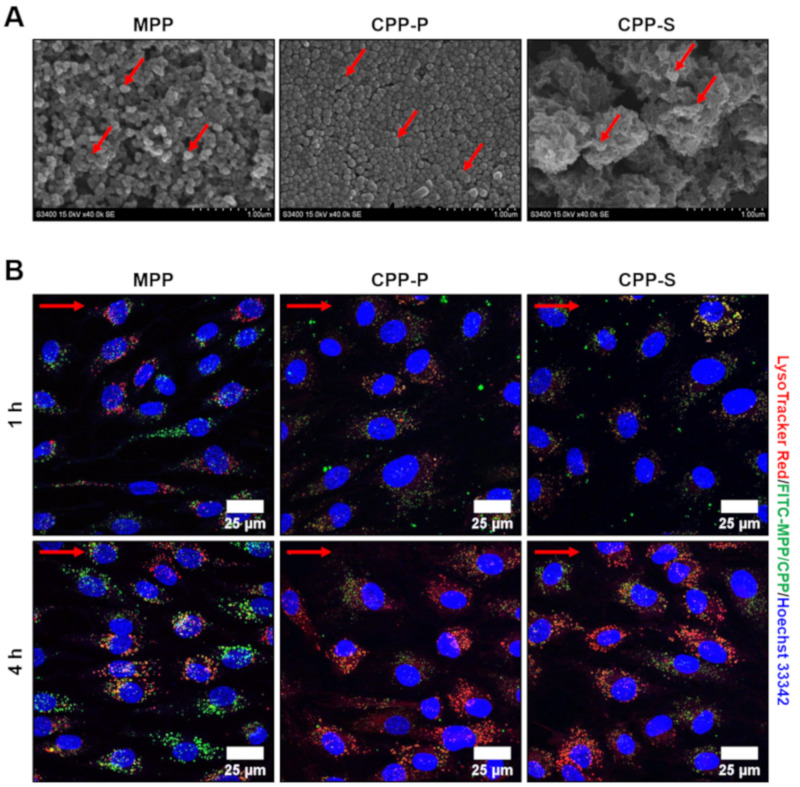
Appearance and internalisation of MPP, CPP-P and CPP-S by primary human ECs. (A) scanning electron microscopy of MPP, CPP-P and CPP-S, representative images, ×40,000 magnification. Red arrows indicate MPPs, CPP-P, and CPP-S; (B) confocal microscopy of HCAEC incubated with FITC-labeled MPP, CPP-P and CPP-S (green colour) for 1 or 4 h and additionally stained by a phago/lysosome-specific dye LysoTracker Red (red colour) along with nuclear counterstaining by Hoechst 33342 (blue colour). Note the internalisation of CPP-P and CPP-S (green dots) and their partial co-localisation with phago/lysosomes (yellow/orange dots) at both 1 and 4 h post treatment. Internalisation of MPPs (green dots) also occurred 1 h post incubation, yet co-localisation with phago/lysosomes (yellow/orange dots) was observed only at 4-h time point. Red arrows indicate the direction of flow. MPP—magnesiprotein particles, CPP-P—primary calciprotein particles, CPP-S—secondary calciprotein particles, HCAEC—human coronary artery endothelial cells, HITAEC—human internal thoracic artery endothelial cells, FITC—fluorescein isothiocyanate.

