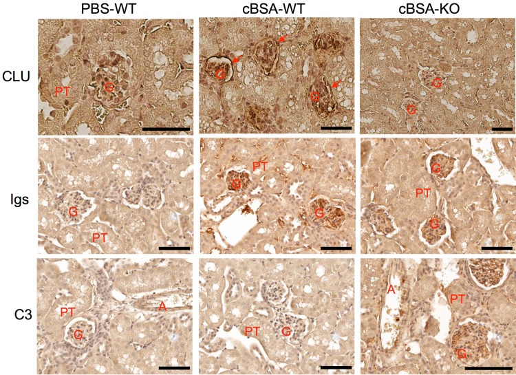
Figure 4.
The glomerular deposit of Igs and C3 in mice after cBSA immunization in CLU-KO mice. The glomerular expression of CLU, Igs or C3 was detected by using immunohistochemical staining. Upper panel: CLU protein. Red arrow: capsular epithelium. Middle panel: Igs. Bottom panel: C3. G: glomerulus, PT: proximal tubule, A: artery. Data are presented in a typical microscopic image of immunohistochemical staining of each target protein. Brown color: positive staining. Scale bar: 80 µm.

