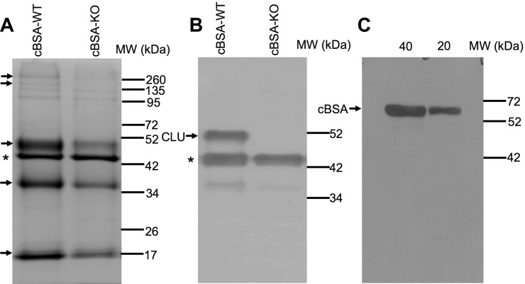
Figure 7.
CLU-bound proteins in the serum of mice after cBSA immunization. Serum CLU and its bound proteins were immunoprecipitated by using anti-CLU antibodies. (A) A typical Coomassie blue staining of fractions of immunoprecipitate in 10% SDS-PAGE. Arrow: the protein bands of interest for LC-MS analysis. (B) Western blot analysis of CLU protein in the immunoprecipitate. * The heavy chain of anti-CLU antibody. (C) Western blot analysis of anti-cBSA IgG in the immunoprecipitate, in which the immunoprecipitates were used as first antibody in the detection of cBSA in the blot. Data present a typical stained PAGE or Western blot of three separate experiments.

