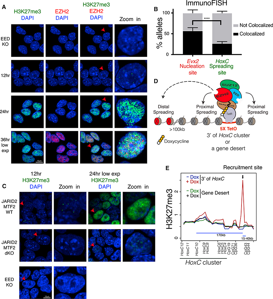
Figure 6. H3K27me3 Domains Initiate within Nuclear Hubs of PRC2 Activity, from which H3K27me3 Spreads in cis and far-cis via Long-Range Interactions.
(A) Immunofluorescence using the indicated antibodies at 0, 12, 24, and 36 hr after EED expression in i-WT-r mESCs. H3K27me3 staining at 36 hr is shown at lower exposures. The rightmost panel is a zoomed-in image of the cell labeled with a red arrow.
(B) Quantification of percentage of alleles colocalized with H3K27me3 foci using immuno-FISH, comparing probes for a nucleation site (Evx2) and a spreading site (HoxC) at 12 hr after EED expression in i-WT-r cells. n for Evx2: 98; n for HoxC: 100. Fisher’s exact test of a combination of two biological replicates: ****p value <0.0001. Error bars represent the SD of the mean.
(C) Immunofluorescence using H3K27me3 antibodies at 12 (left) and 24 (right) hr after rescue of EED expression in i-WT-r mESCs with the indicated genotypes. H3K27me3 staining at 24 hr is shown at lower exposures. The rightmost panel is a zoomed-in image of the cell labeled with a red arrow. Staining of EED KO cells (0 hr, without 4-OHT) is shown at bottom left as a control.
(D) Scheme for ectopic targeting of PRC2 to test proximal and distal spreading of its activity using the Tet repressor and Tet operator system in i-WT-r cells (see STAR Methods for details).
(E) Average scaled ChIP-seq read density of H3K27me3 plotted near the recruitment site using a 10-kb window centered around H3K27me3 peaks found in WT cells, before (–Dox) and 24 hr after (+Dox) induction of PRC2 recruitment downstream of the HoxC cluster and EED expression in i-WT-r mESCs. HoxC10 spatially interacted with the recruitment site downstream of the HoxC cluster and with the genes indicated and with CpGs, as determined by 4C-seq.

