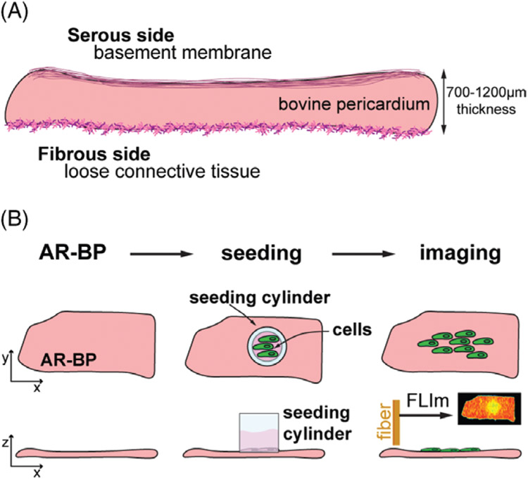
FIGURE 1.
Scaffold and experimental timeline. (A) Sketch of the scaffold properties. Bovine pericardium (1-2 mm in thickness) is composed of structural proteins organized in a basement membrane on the serous side, and as loose connective tissue on the fibrous side, providing 2 different extracellular matrix niches for re-seeded cells. (B) The experimental design consists on 3 main steps: (1) antigen removal of the scaffold to generate antigen removed bovine pericardium (AR-BP), (2) seeding the scaffold with either human aortic endothelial cells (hAECs) or human mesenchymal stem cells (hMSCs) and (3) imaging the recellularization process through fiber optic fluorescence lifetime imaging (FLIm) over 7 days after seeding

