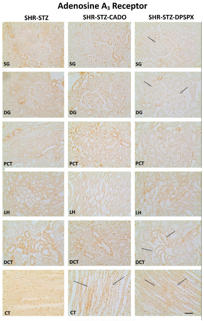
Figure 5.
Representative photomicrographs of the immunoreactivity against the adenosine A3 receptors in the superficial (SG) and deep glomeruli (DG), proximal convoluted tubule (PCT), distal convoluted tubule (DCT), loop of Henle (LH), and collecting tubule (CT) of SHR-STZ (left panel), SHR-STZ + CADO (middle panel), and SHR-STZ + DPSPX (right panel) animals. Open arrows: evidence less pronounced immunoreactivity than that exhibited by SHR-STZ. Bars: 20 µm.

