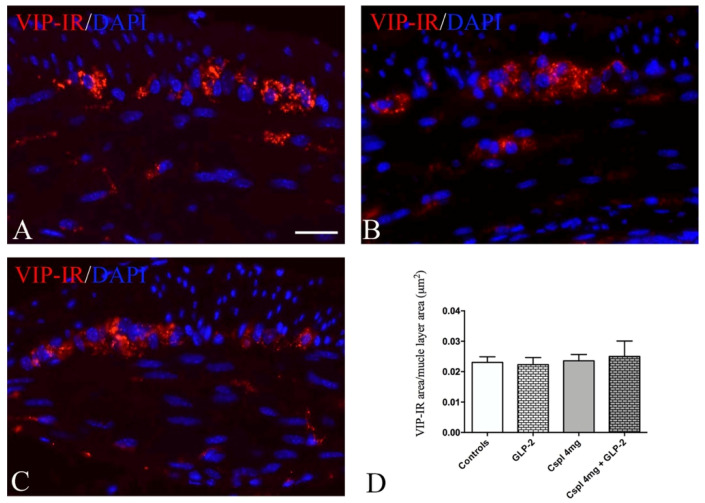
Figure 8.
VIP labeling in muscle wall (A–C). Controls (A), cisplatin (B) and cisplatin + [Gly2]GLP-2 (C) group. VIP labeling was detected as small granules located within the ganglia and muscle layers. DAPI labeling stained the nucleus. Scale bar = 20 μm. Quantitation of VIP-IR structures (D). The assessment of VIP expression was performed considering the whole cross-section. Data are expressed as mean ± SEM. One-way ANOVA test, post hoc Newman Keuls’. No significance. n = 4–6 for each group.

