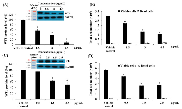
Figure 10.
Effect of F-EtOAc and compound 1 at various concentrations on KG-1a cells. (A) The level of WT1 protein following treatment with F-EtOAc for 48 h. Protein levels were evaluated using western blot and analysed using a scanning densitometer. The levels of WT1 were normalised using GAPDH protein levels. (B) Total cell number following treatment with F-EtOAc for 48 h. Total cell numbers were determined via the trypan blue exclusion method. (C) The level of WT1 protein after treatment with compound 1 for 72 h. Protein levels were evaluated using Western blotting and analysed using a scanning densitometer. The levels of WT1 were normalised using GAPDH protein levels. (D) Total cell number following treatment with compound 1 for 72 h. Total cell numbers were determined via the trypan blue exclusion method. Each bar represented mean ± SD of three independent experiments performed in triplicate. Asterisks (*) denote significant differences from the vehicle control (* p < 0.01).

