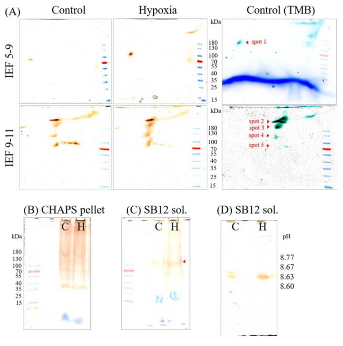
Figure 4.
Abundance of guaiacol peroxidases under hypoxia-stress. Plasma membranes (250 µg total protein) of control (C) and 24 h hypoxia-stressed (H) maize roots were solubilized with 8% CHAPS for 1 h. The supernatant was separated on (A) native IEF gels pH 5–8 and pH 9–11 followed by 4–18% non-denaturing polyacrylamide gels in second dimensions. The remaining pellet was either separated on 4–18% non-denaturing gels (B) or solubilized with SB12 (protein-detergent ratio of 1:7) for 1 h. The SB12 supernatant (sol.) was loaded on 4–18% non-denaturing gels (C) and IEF gels pH 9–11 (D). Class III peroxidases were visualized by staining with hydrogen peroxide and guaiacol (orange color). No or only weak signal could be determined in the pellet after SB12 solubilization (data not shown). For MS analyses, gels were stained with TMB (blue color). The protein spots and bands, indicated with red arrows, were identified by mass spectrometry. Molecular weight in kDa was determined using a protein standard (PageRuler Prestained Protein Ladder, Thermo Scientific, Waltham, MA, USA). For further details, see the text. SB12, n-dodecyl-N, N-dimethyl-3-ammonio1-propanesulfonate; CHAPS, 3-[(3-Cholamidopropyl)dimethylammonio]1-propanesulfonate; TMB, 3,3′,5,5′-Tetramethylbenzidine.

