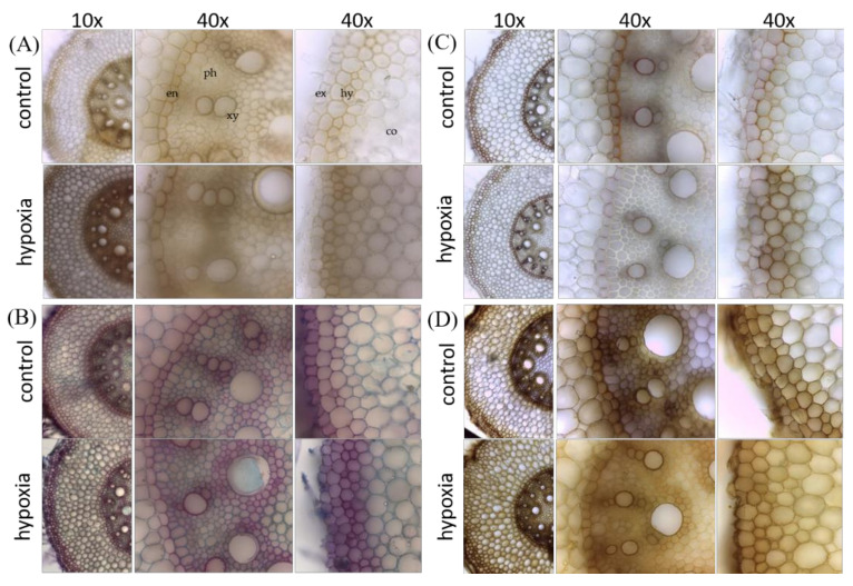
Figure 7.
Visualization of lignin, its precursors, and cellulose in the mature zone of primary root cross-sections from control and hypoxia-stressed maize plants. Control and 24 h hypoxia-stressed maize roots were cross-sectioned by hand and analyzed with (A) Mäule staining for syringyl-rich polyphenols (deep-red color), (B) fuchsin, chrysoidin, and Astra blue (FCA) staining for lignified cells (red color) and non-ligneous cells (blue color), (C) Phloroglucinol staining for lignified cells (pink color), and (D) chlorine–zinc–iodine staining for cellulose (violet color). For detailed explanation of the colours, see methods. Co, cortex; en, endodermis; ex, exodermis; hy, hypodermis; ph, phloem; xy, xylem.

