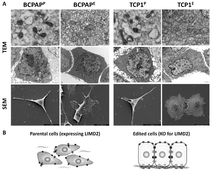
Figure 4.
(A) Representative images of transmission electron microscopy (TEM) and scanning electron microscopy (SEM). Parental cells (BCPAPP and TPC1P) presented loss of apical-basal polarity and concentration of mitochondria in one pole of the cells. The presence of small and circular mitochondria suggests mitochondrial fission. Vacuoles and mitochondrial fission are showed (arrow). Remarkably, the apical-basal polarity and mitochondria morphology are somewhat restored in LIMD2-edited cells (BCPAPE and TPC1E); (B) Schematic model of parental (expressing LIMD2) and edited cells (complete KO of LIMD2).

