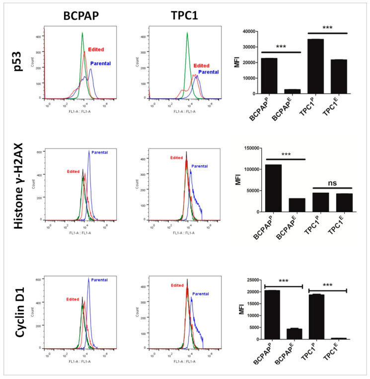
Figure 10.
Results of immunodetection of p53, histone γ-H2AX, and cyclin D1 by flow cytometry. Results show a significant downregulation of all analyzed proteins in both edited cells. Flow cytometry analyses show cells not incubated with primary and secondary antibodies (histogram in black), cells exclusively incubated with secondary antibody (histogram in green), and cells incubated with both primary and secondary antibodies (histogram in red). There were 10,000 events analyzed in duplicate. *** p < 0.001, non-significant statistical difference (ns).

