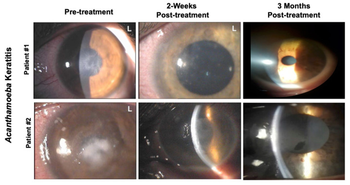
Figure 2.
Representative pictures of two patients affected by Acanthamoeba Keratitis. Pictures were taken at different time points pre- and post-treatment with a solution of CHX (chlorhexidine) 0.02% and VE-T-GS 0.2%. Patient #1: cornea shows epithelial irregularity and punctate epithelial lesions before treatment. Patient #2: cornea shows a gray-white diffuse stromal infiltrate positioning ring0shaped with stromal thinning prior the treatment.

