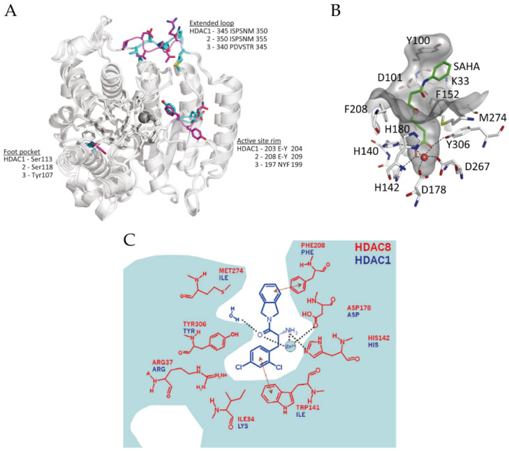
Figure 2.
(A) Superposition of the structures of HDAC1–3 (PDB (Protein Data Bank) codes: 5ICN, 4LY1 and 4A69). Significant residue differences are highlighted in cyan (HDAC1 and 2) and magenta (HDAC3). Adapted with permission from Millard 2017 [15]. (B) The active site of HDAC8 (PDB code: 4QA2). Adapted with permission from Chakrabarti 2015 [17]. (C) The foot pocket of HDAC8. Prominent amino-acid side-chain differences between HDAC8 and HDAC1 in the foot pocket are shown. Adapted with permission from Whitehead 2011 [64].

