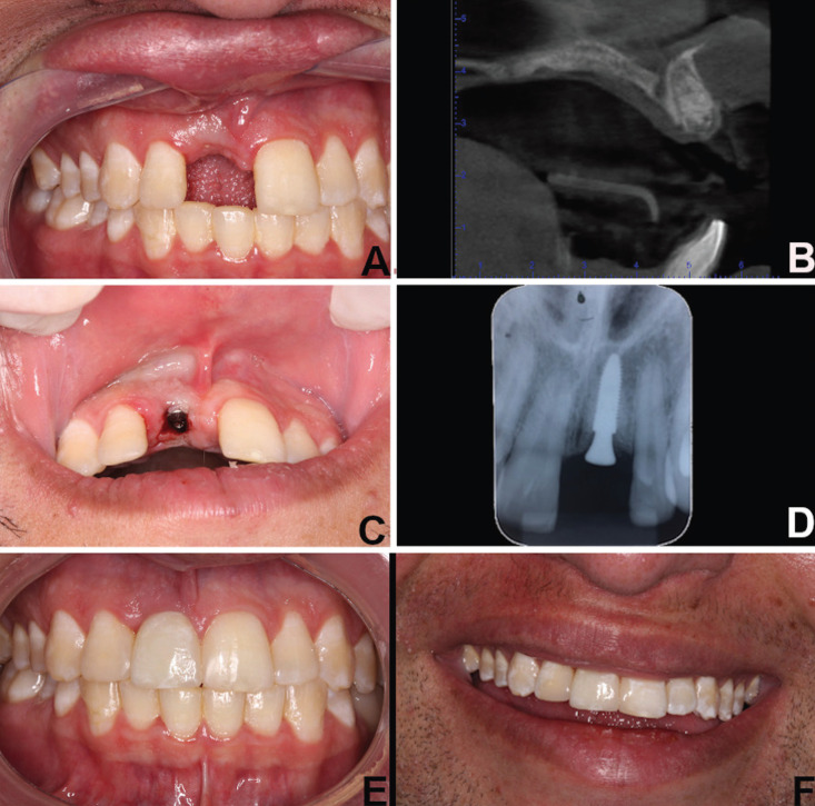
Figure 2.
A) Clinical examination after suture removal. B) Axial section of cone beam computed tomography after the healing and complete osseointegration of the grafted biomaterial. C) Implant installation surgery and healing abutment placed simultaneously at the surgical procedure. D) Intraoral periapical radiographs showing implant osseointegrated. Intraoral E) and F) extraoral examination showing clinical appearance after permanent ceramic crown on tooth 11 and restoration on tooth 12 and 21.

