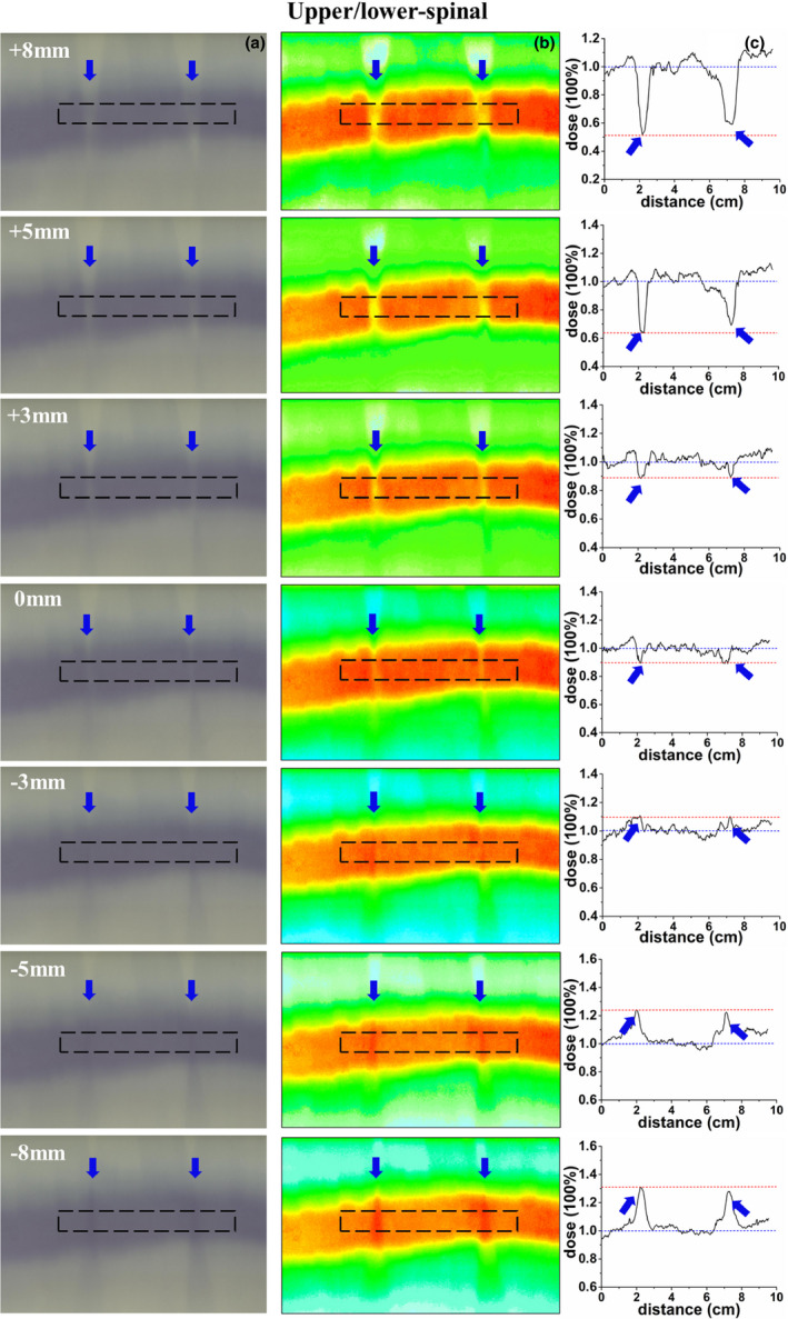
Fig. 7.

Verification of the upper/lower‐spinal field junction; (a) an irradiated radiochromic film, (b) dose distribution of the radiochromic film, (c) dose profile along the dotted line, the dose profile was normalized to the prescription dose, and the blue arrow represent the hot/cold peaks of dose in the figures.
