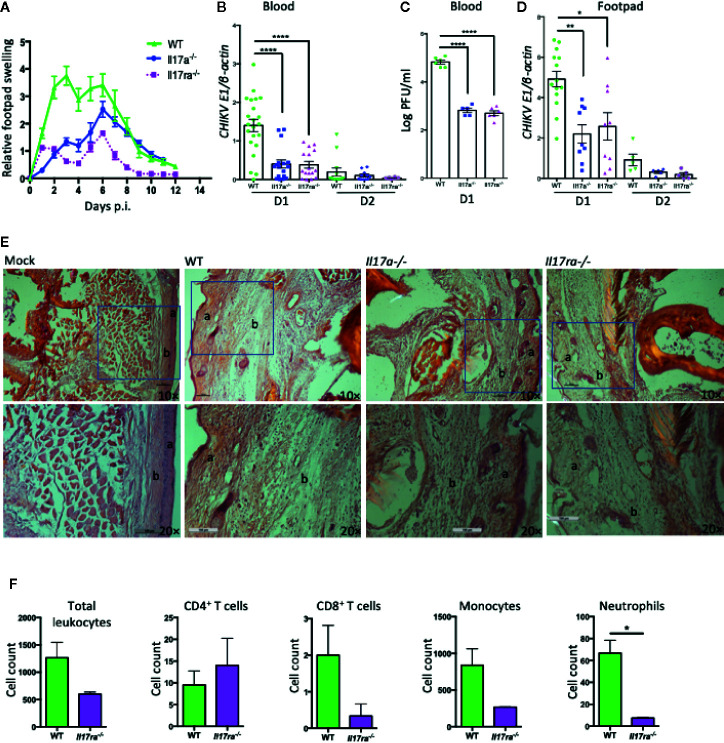
Figure 2.
Il17a−/− and Il17ra−/− mice are more resistant to CHIKV infection. Seven to eight weeks old WT, Il17a−/− and Il17ra−/− mice were infected with 1 × 10⁵ PFU of CHIKV via footpad. (A) Swelling of injected footpads (peri-metatarsal area) of mice is presented as a relative increase in swelling that was calculated by measuring the thickness and width of an inoculated footpad. (B) Blood was collected on days 1 (D1) and D2 p.i., and QPCR was performed to measure the copies of CHIKV E1 and cellular β-actin transcripts. Viral burden was expressed as a ratio of copy number of CHIKV E1 to cellular β-actin transcripts and compared between WT (n = 22) and Il17a−/− (n = 17) or Il17ra−/− (n = 16) mice. (C) Plaque assay was performed to measure the viral load in the blood samples of D1 from (B). (D) Inoculated footpads of WT (n = 14) and Il17a−/− (n = 9) or Il17ra−/− (n = 9) were collected on D1 and D2 p.i., and the viral burden was quantified by QPCR after normalization to cellular β-actin. (E) Representative H&E stained histological images (10×, upper row and 20×, lower row) of peri-metatarsal area of the inoculated foot tissue (n = 5 mice/group) on D6 p.i. The layer of epidermis (a) and dermis (b) are labeled. (F) Seven to 8 weeks old WT (n = 4) and Il17ra−/− mice (n = 3) were infected with 1 × 10⁵ PFU of CHIKV via footpad. Footpad cells were isolated at D6 p.i., and quantified by flow cytometry after probing with antibodies against CD45, CD4, CD8, CD11b, and Ly6G. The cell counts for positive cells within the gated population are shown for WT and Il17ra−/− mice. The results are expressed as mean ± SEM (A–D, F) or a representative image (E) that represents at least three independent experiments. The data were analyzed by a two-tailed Student t-test (A–D, F). *, **, and **** denote p < 0.05, p < 0.01, and p < 0.0001 respectively, when compared to WT controls).

