FIGURE 2.
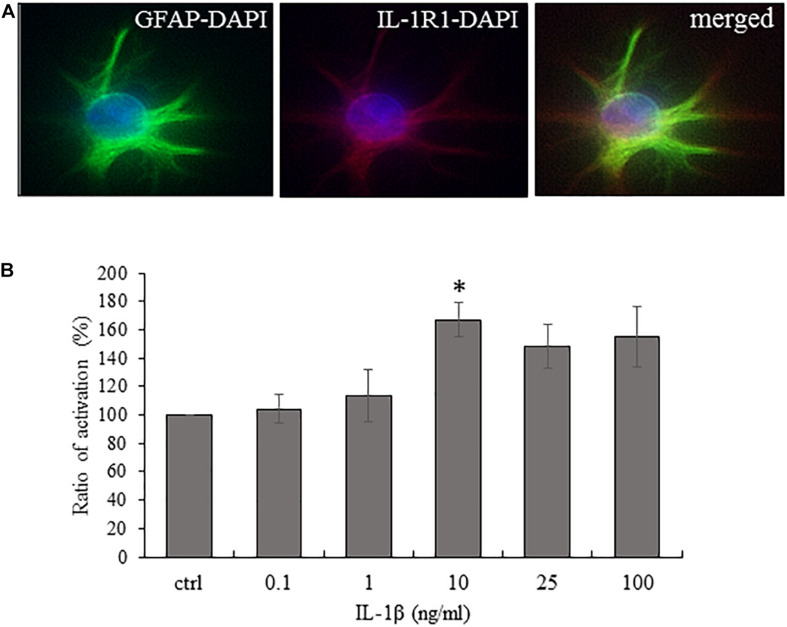
Cultured spinal astrocytes express IL-1R1 and their activity is significantly increased upon IL-1β stimulation. (A) Cultured spinal astrocytes express the ligand binding unit or IL-1 receptor (IL-1R1). Micrographs of fluorescent images illustrating co-localization between GFAP astrocytic marker (a,c; green) and IL-1R1 (b,c; red). Panel a–c represent control cultures. Mixed colors (yellow) on the superimposed image (c) indicate double labeled structures. On all images DAPI was used to label cell nuclei (blue). (B) Dose-dependent (1–100 ng/ml) enhancement of astrocytic activity by IL-1β was determined by MTT assay after 24 h of treatment. Quantification of MTT activity is presented as fold change over the control cells in percentage. Data are shown as mean ± SEM of three independent experiments in duplicate assay (ANOVA Repeated Measures, followed by Tukey’s pairwise comparison ∗p < 0.05 versus control group).

