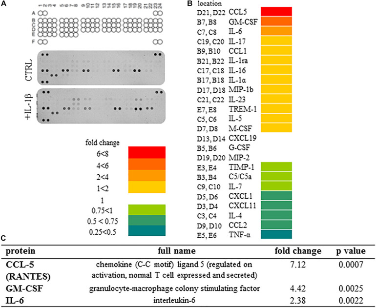
FIGURE 3.
Secretome profile of IL-1β treated spinal astrocytes reveals the expression of 24 cytokines and chemokines. (A) Coordinates of cytokines on array membranes and developed membranes. The Proteome Profiler array used in the experiments allowed the detection of 40 cytokines, chemokines and other soluble factors. Upper panel shows array coordinates. Middle panel shows expression of cytokines/chemokines in the supernatant of unstimulated spinal astrocytes, and lower panel shows expression of cytokines/chemokines by IL-1β stimulated astrocytes. (B) Heat map analysis of spinal astrocytic cytokine profile after 24 h of IL-1β stimulus. Left column shows array coordinates of cytokines/chemokines in the middle column and right column shows the fold change of their amount. (C) List of cytokines/chemokines which are significantly overexpressed in the supernatant of spinal astrocyte cultures upon IL-1β stimulation.

