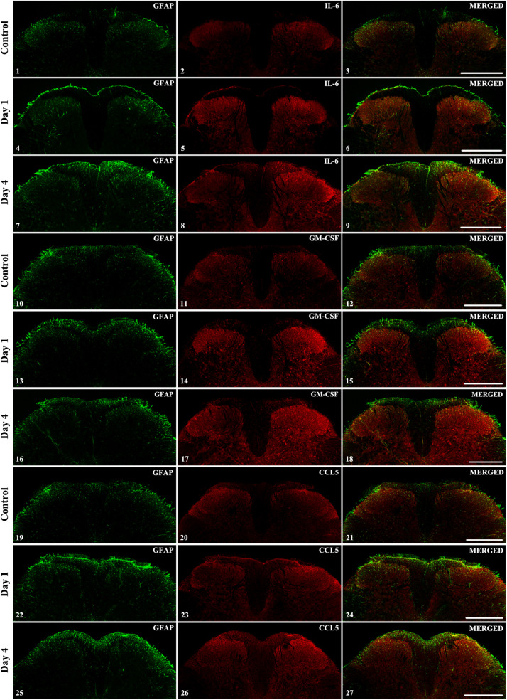
FIGURE 6.
The spinal expression of IL-6, GM-CSF, and CCL5 cytokines during the course of CFA-evoked pain. Representative 1 μm thick confocal images illustrating the co-localization between immunolabeling for IL-6 (red; 6.2, 6.5, 6.8), GM-CSF (red; 6.11, 6.14, 6.17) CCL5 (red; 6.20, 6.23, 6.26) and the immunoreactivity of astrocytes (GFAP, green; first column of the figures) in the superficial spinal dorsal horn. Mixed colors (yellow) on the superimposed images (last column of the figures) indicate double-labeled structures. For each cytokine the first row of the images are taken from control samples, whereas the second row of images represents the first day after CFA injection and the third row shows the spinal expression of the cytokines on post-injection day 4. Increased labeling of the cytokines was apparent on the ipsi-lateral (right side) of the spinal cords on post-injection day 4 (panels 6.8, 6.17, and 6.26). In each case scale bars: 500 μm.

