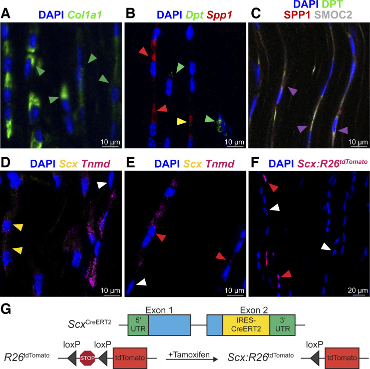
Fig. 3.
RNA in situ hybridization and histology of tendon markers. RNA in situ hybridization (A, B, D, and E) and protein fluorescence and immunofluorescence (C and F) of mouse Achilles tendons. A: Col1a1 RNA, green; green arrowheads exemplify Col1a1+ fibroblasts. B: mRNA: Dpt, green; Spp1, red. Green arrowheads exemplify Spp1−Dpt+ fibroblasts; red arrowheads exemplify Spp1+Dpt− fibroblasts; yellow arrowheads exemplify Spp1+Dpt+ fibroblasts. C: protein: DPT, green; osteopontin, red; and SMOC2, white. Magenta arrowheads exemplify overlap between DPT, SPP1, and SMOC2 protein. D–E: RNA: Scx, yellow; Tnmd, violet. Red arrowheads exemplify Scx−Tnmd+ fibroblasts; yellow arrowheads exemplify Scx+Tnmd+ fibroblasts; and white arrowheads exemplify Scx−Tnmd− fibroblasts. F: tdTomato protein identifies scleraxis-expressing cells (red arrowheads) compared with non-scleraxis expressing cells (white arrowheads). Nuclei visualized with DAPI. G: genetics of scleraxis lineage-tracing mice. Representative images of 4 mice.

