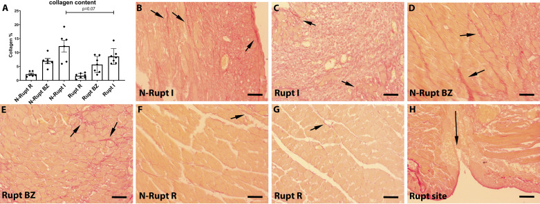
Fig. 6.
Rupture is associated with a trend toward reduced collagen deposition in the healing infarct. A: quantitative analysis using a machine learning approach showed that collagen content in the infarct (I) was lower in mice dying of rupture (Rupt) compared with animals dying without rupture (N-rupt). However, the difference did not reach statistical significance (P = 0.07, n = 6–7/group). No significant differences were noted in the border zone (BZ) and in the remote remodeling myocardium (R). B–H: representative images show labeling of collagen fibers using Picrosirius red staining (short arrows). H: collagen was absent in the rupture site (long arrow). Time points studied histologically were comparable between groups, as there was no significant difference in the time of death (rupture group: 5.0 ± 0.53 days, n = 7; no rupture group: 5.16 ± 0.4, n = 6). Scale bar, 50 μm.

