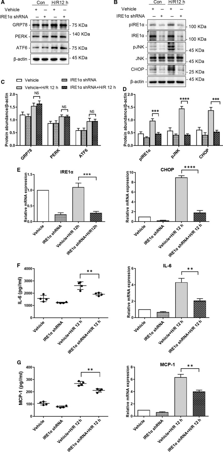
FIGURE 2.

Depletion of IRE1α reduces ER stress and the production of inflammatory cytokines in HK‐2 cells following hypoxia‐reoxygenation (H/R). HK‐2 cells depleted of IRE1α and vehicle‐infected HK‐2 cells were cultured under normal conditions or subjected to H/R (4 h of hypoxia followed by 12 h reoxygenation). Representative western blots of ER stress markers (A) upstream (GRP78, PERK, ATF6) and (B) downstream (pJNK, CHOP) of IRE1α; β‐actin was used as the loading control. Quantitative analysis of western blots of ER stress markers (C) upstream (GRP78, PERK, ATF6) and (D) downstream (p‐JNK, CHOP) of IRE1α. E, qRT‐PCR analysis of IRE1α, CHOP and ACTB (internal control). F, IL‐6 secretion to culture supernatant, and qRT‐PCR analysis of IL‐6 and ACTB (internal control). G, MCP‐1 secretion to the culture supernatant, and qRT‐PCR analysis of MCP‐1 and ACTB (internal control). Con: HK‐2 cells without H/R; H/R 12 h:HK‐2 cells with 4 h of hypoxia followed by 12 h reoxygenation; IRE1αShRNA: IRE1α shRNA‐infected HK‐2 cells; Vehicle: vehicle‐infected HK‐2 cells. (NS: not significant, **P < .01, ***P < .001, ****P < .0001)
