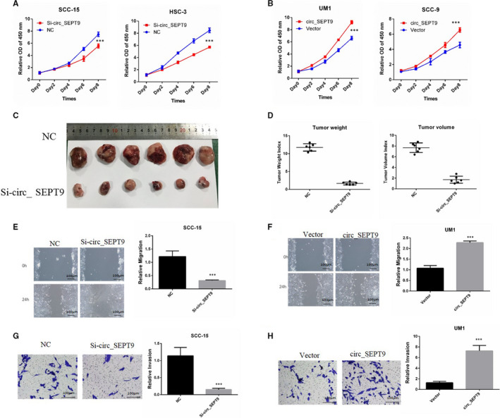
FIGURE 3.

circ_SEPT9 promotes the progression of OSCC cells. A, CCK8 was performed to detect the growth rates of SCC‐15 and HSC‐3 with transfection of si_circSEPT9. B, CCK8 was used to detect the growth rates of UM1 and SCC‐9 with transfection of circ_SEPT9 overexpression plasmids. C, 2.5 × 10⁶ si‐circ_SEPT9 and NC SCC‐15 cells were subcutaneously injected into the flank of the nude mice (n = 6). Photographs of tumours derived from nude mice were shown. D, Tumour weight and tumour volume were analysed and shown. E, Wound assay was performed in SCC‐15 cells. Photographs were taken by microscope at 0 and 24 h. The scale bar is 100 μm. F, Wound assay was performed in UM1 cells. Photographs were taken by microscope at 0 and 24 h. The scale bar is 100 μm. G, Transwell assay was performed in SCC‐15 cells. Invasive cells were stained and counted. The scale bar is 100 μm. H, Transwell assay was performed in UM1 cells. Invasive cells were stained and counted. The scale bar is 100 μm
