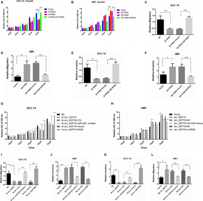
FIGURE 7.

circ_SEPT9/miR‐1225/PKN2 contribute to the progression of OSCC cells. A, CCK8 was performed to detect the growth rate of SCC‐15. B, CCK8 was performed to detect the growth rate of UM1. C, Wound assay was performed in SCC‐15 cells. Photographs were taken by microscope at 0 and 24 h. D, Wound assay was performed in UM1 cells. Photographs were taken by microscope at 0 and 24 h. E, Transwell assay was performed in SCC‐15 cells. F, Transwell assay was performed in UM1 cells. G, SCC‐15 cells were transfected with indicated plasmids. CCK8 was performed to detect the growth rate. H, UM1 cells were transfected with indicated plasmids. CCK8 was performed to detect the growth rate. I, Wound assays were performed to detect the migration ability of SCC‐15. J, Wound assays were performed to detect the migration ability of UM1. K, Transwell assays were used to detect the invasion ability of SCC‐15. L, Transwell assays were used to detect the invasion ability of UM1
