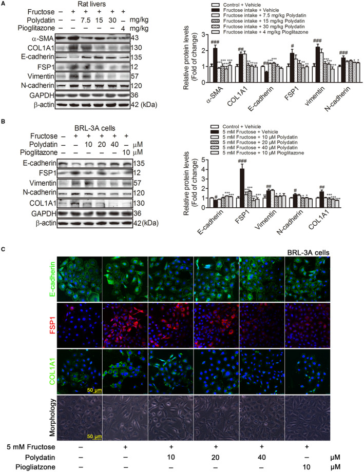
FIGURE 2.

Polydatin attenuates fructose‐induced EMT in liver fibrosis of rats and BRL‐3A cells. (A) Western blot analysis of α‐SMA, COL1A1, E‐cadherin, FSP1, vimentin and N‐cadherin protein levels in rat livers. (B) Western blot analysis of E‐cadherin, FSP1, vimentin, N‐cadherin and COL1A1 protein levels (48 h) in BRL‐3A cells cultured with or without 5 mM fructose in the presence or absence of polydatin (10, 20 and 40 μM) or pioglitazone (10 μM). GAPDH and β‐actin were as internal control. (C) Images of BRL‐3A cells labelled with antibodies specific for E‐cadherin (green), FSP1 (red), COL1A1 (green) as well as morphology. Each value is shown as mean ± SEM (n = 4‐6). # P < .05, ## P < .01, ### P < .001 compared with the normal control; *P < .05, **P < .01, ***P < .001 compared with the fructose control. FSP1, fibroblast specific protein l; α‐SMA, α smooth muscle actin; COL1A1, collagen 1
