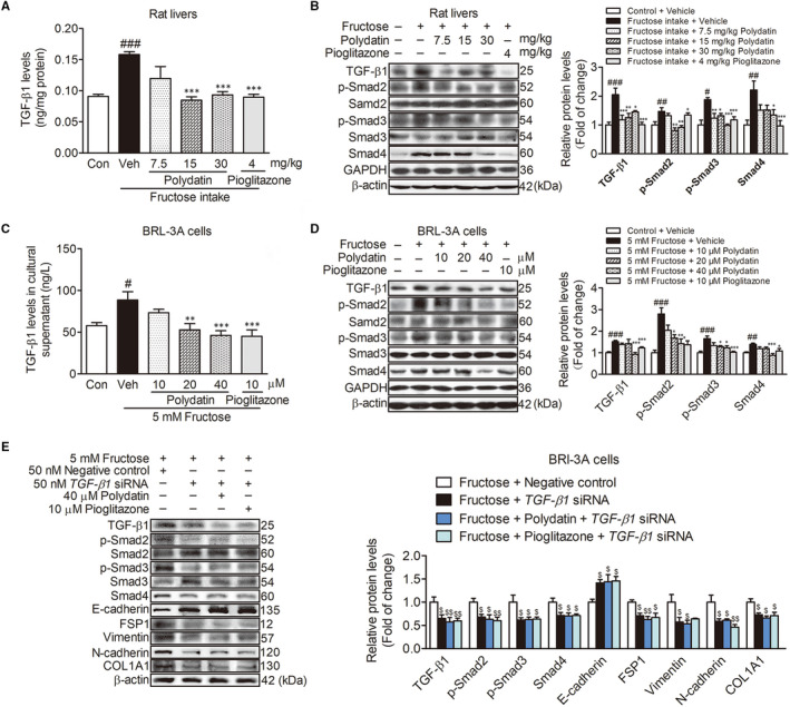
FIGURE 3.

Polydatin suppresses the activation of TGF‐β1/Smad signalling to attenuate fructose‐induced EMT. (A) The liver concentration of TGF‐β1 in the rats was measured with ELISA kit. (B) Western blot analysis of TGF‐β1, p‐Smad2, p‐Smad3 and Smad4 protein levels. (C) TGF‐β1 secretion levels in BRL‐3A cell supernatant were detected with ELISA kit. (D) Western blot analysis of TGF‐β1, p‐Smad2, p‐Smad3 and Smad4 protein levels in BRL‐3A cells (48 h). (E) Western blot analysis of TGF‐β1 (24 h), p‐Smad2, p‐Smad3, Smad4, E‐cadherin, FSP1, vimentin, N‐cadherin and COL1A1 (48 h) protein levels in transfected with 50 nM TGF‐β1 siRNA or NC BRL‐3A cells treated with fructose in the presence or absence of 40 μM polydatin or 10 μM pioglitazone. GAPDH and β‐actin were as internal control. Each value is shown as mean ± SEM (n = 4‐6). # P < .05, ## P < .01, ### P < .001 compared with the normal control; *P < .05, **P < .01, ***P < .001 compared with the fructose control; $ P < .05, $$ P < .01 compared with the fructose‐negative control
