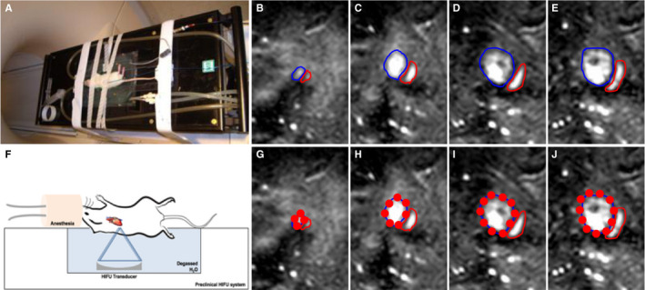
Figure 1.

MR‐guided pFUS. A, Rats placed on a MR‐compatible pre‐clinical focused ultrasound system. F, Illustration of experimental setup of pFUS treatment to the rat heart. Left side of the rat chest was submerged in degassed water to pose the heart perpendicular to the focused ultrasound transducer. B‐E, Sequential T2w coronal MR images were acquired at 1 mm slice thickness for pFUS to target the left ventricle of the rat heart. Blue outline area represents the left ventricle, and red outline area represents the right ventricle. G‐J, pFUS targeting spots in red circle based on the MR guidance images
