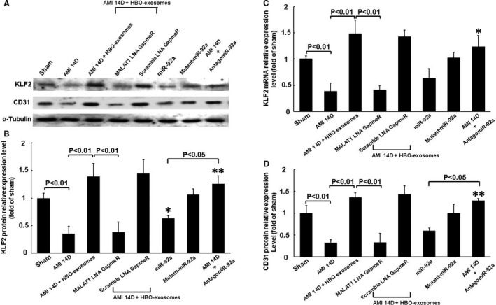
Figure 3.

MALAT1 and miR‐92a mediate myocardial KLF2 and CD31 expression in AMI rats. A, Representative Western blot analysis for KLF2, CD31 and α‐tubulin protein expression in the rat left ventricular myocardium. MALAT1 LNA GapmeR and miR‐92a expression vector were transfected into left ventricular myocardium using a low pressure‐accelerated gene gun. B, Quantitative analysis of KLF2 protein levels. The values of protein expression from left ventricular myocardium after AMI were normalized to α‐tubulin measurement and then expressed as a ratio of normalized values to protein in the sham group. *P < .05 vs sham. **P < .01 vs AMI 14 d (n = 5 per group). C, Quantitative real‐time PCR of KLF2 mRNA expression in rat left ventricular myocardium. *P < .01 vs AMI 14 d (n = 5 per group). D, Quantitative analysis of CD31 protein levels. The values of protein expression from left ventricular myocardium after AMI were normalized to α‐tubulin measurement and then expressed as a ratio of normalized values to protein in sham group. *P < .05 vs sham. **P < .01 vs AMI 14 d (n = 5 per group)
