
Figure 4.
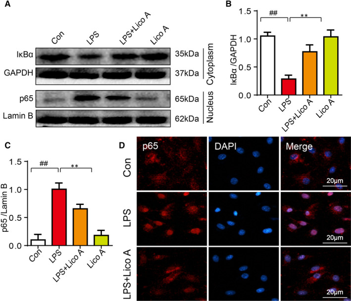
Effects of Lico A on LPS‐stimulated NF‐κB activation in mouse chondrocytes. The chondrocytes were treated with or without LPS (1 μg/mL) and Lico A for 6 h, and then following treatment of ATP (5 mmol/L) for 30 min after incubation overnight. The protein expression levels of p65 and IkBα treated as above were visualized by Western blot (A) and are quantified in (B‐C). The nuclear translocation of p65 was detected by immunofluorescence combined with DAPI staining for nuclei (D).Values represent the averages ± SD Significant differences between different groups are indicated as ## P < 0.01, vs control group; **P < 0.01, vs LPS alone treatment group, n = 5