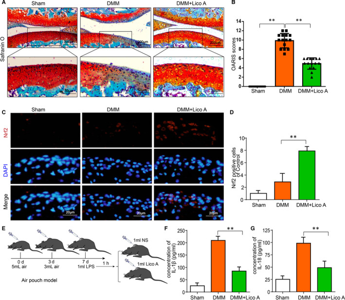
Figure 6.

Lico A mitigates OA progression and enhances the expression of Nrf2 in a mouse DMM model. Safranin O staining was used to assess histomorphometric differences between experimental groups at 8 wk post‐surgery (A). Histological analysis of OA was evaluated by Osteoarthritis Research Society International (OARSI) scores (B). Nrf2 (C) was detected by immunofluorescence combined with DAPI staining. The fluorescence intensity of Nrf2 were analysed by ImageJ (D). Air pouch mouse model was used to assess the effect of Lico A on the production of IL‐1β and IL‐18 stimulated by LPS in vivo (E). The production of IL‑1β and IL‐18 in mice chondrocytes treated as above were measure by ELISA (F‐G).Values represent the averages ± SD Significant differences between different groups are indicated as **P < 0.01 vs the Sham group and **P < 0.01 vs the DMM group, n = 15
