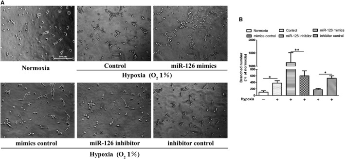
Figure 6.

miR‐126‐mediated tube formation of HUVECs. Cells were treated 4 hours in normoxic condition (21% O2, 5% CO2) or hypoxic condition (1% O2, 5% CO2 and 94% N2). (A) Images of tube formation assay in all groups. (B)The branched numbers analysed by ImageJ (normoxia group was set to 100%). Values are expressed as mean ± SD (n = 3/group), *P < .05; **P < .01
