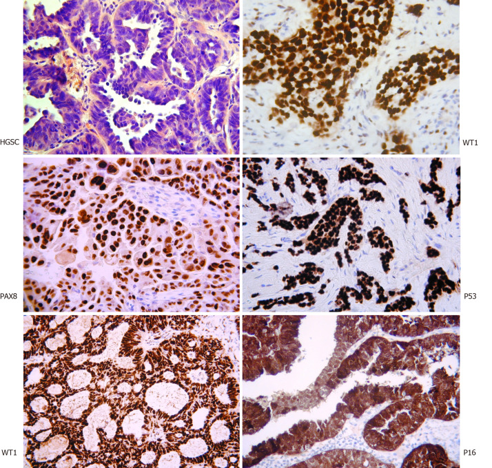
Figure 1.
Histopathological assessment of high-grade serous carcinoma. The classical appearance on hematoxylin and eosin with intermediate sized tumor cells, marked nuclear atypia, and necrotic areas. Immunostaining with WT1, PAX8, P16, and P53 assist with the diagnosis. Interestingly WT1 staining helps discriminate between high-grade serous carcinoma and pseudo-endometrioid (bottom left) (Original figure, images courtesy of Professor McCluggage WG).

