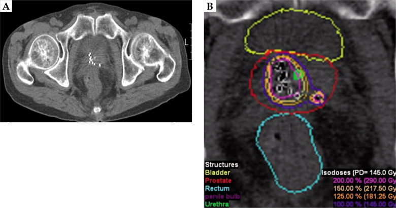
Fig. 2.
Example images of focal zonal type brachytherapy of a 69-year-old patient with a prostate-specific antigen concentration of 4.8 ng/ml. Digital rectal examination showed no abnormalities. A 12-core transrectal ultrasound-guided prostate biopsy revealed Gleason 3 + 3 tumor in four cores in the right lobe of prostate. A) Post-implant axial computed tomography image, B) post-implant dosimetry

