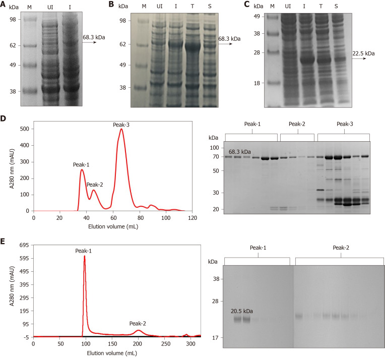
Figure 1.
Expression and purification of full-length His6-NS3 and non-structural protein 4a-non-structural protein 3 protease domain. A: Analysis of the full-length His6–NS3 protein (68.3 kDa) expressed in Escherichia coli BL21 (DE3) using pET11a-His6-NS3. Lane M: SeeBlueTM Pre-Stained Protein Marker (LC5625); Lane UI: Uninduced BL21 (DE3) cells and Lane I: Induced BL21 (DE3) cells; B: Analysis of soluble and insoluble fractions of the His6–NS3 protein expressed in E. coli BL21-CodonPlus (DE3)-RIL cells using pET11a-His6-NS3. Lane M: Invitrogen Cat 1891868 See Blue® Plus2 Pre-Stained ladder; Lane UI: Uninduced cells; Lane I: Induced cells; Lane T: Total cell lysate of induced cells; Lane S: Soluble fraction of lysed cells; C: Analysis of soluble and insoluble fractions of the His7–non-structural protein 4a-non-structural protein 3 (NS4A-NS3) protease domain expressed in E. coli BL21 (DE3) cells using pET11a-His7-NS4A-NS3. Lane M: SeeBlueTM Pre-Stained Protein Marker (LC5625); Lane UI: Uninduced cells; Lane I: Induced cells; Lane T: Total cell lysate of induced cells; Lane S: Soluble fraction of lysed cells; D: Analysis of the Ni-NTA His6–NS3 protein by gel filtration, presence of proteins in peak 1, 2 and 3 is analyzed by sodium dodecyl sulfate polyacrylamide gel electrophoresis; and E: Analysis of the native NS4–NS3 protease domain by gel filtration, presence of proteins in peak 1 and 2 is analyzed by sodium dodecyl sulfate polyacrylamide gel electrophoresis.

