
Figure 5.
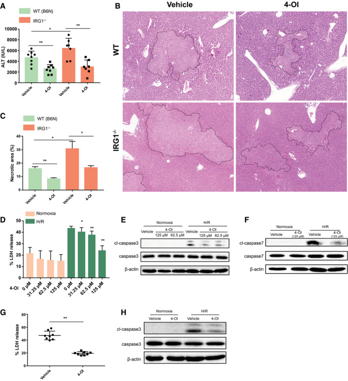
Itaconate ameliorates hepatic I/R injury and rescues hepatocytes from H/R. WT (C57BL/6NJ, B6N) and IRG1−/− mice were subjected to ischemia for 1 hour and then reperfusion for 6 hours. 4‐OI or vehicle control (25 mg/kg body weight) was injected intraperitoneally 2 hours before hepatic I/R and at the time of reperfusion. (A) Serum ALT levels in each group of mice after 1‐hour ischemia and 6‐hour reperfusion; n = 7‐8 in each group. (B) Representative liver H&E staining (original magnification ×20) from each group of mice after 1‐hour ischemia and 6‐hour reperfusion. (C) Dotted lines in liver H&E staining indicate measured areas of necrosis, quantified in bar graph; n = 3 in each group. Primary hepatocytes isolated from WT mice were pretreated with 4‐OI or vehicle control 1 hour prior to normoxia or 10‐hour hypoxia and 10‐hour reoxygenation (H/R). (D) Cell death was assessed by LDH release from hepatocytes after normoxia or H/R; n = 8 for each group. (E,F) Western blot for apoptosis markers (cleaved caspase‐3, cleaved caspase‐7, etc.) in whole‐cell lysates from hepatocytes after normoxia or H/R. Primary hepatocytes isolated from IRG1−/− mice were pretreated with 4‐OI (125 µM) or vehicle control 1 hour prior to normoxia or 10‐hour hypoxia and 10‐hour reoxygenation (H/R). (G) Cell death was accessed by LDH release from hepatocytes after H/R; n = 8 for each group. (H) Western blot for cleaved caspase‐3 and caspase‐3 in whole‐cell lysates from hepatocytes after normoxia or H/R. Images are representative of data from multiple mice per experimental group. Data are presented as means ± SD. *P < 0.05, **P < 0.01. Abbreviation: cl, cleaved.