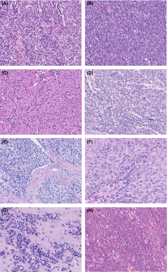
Figure 1.

Examples of thymomas from the thymoma panel; cases A–D were scored with high consensus rates, and for cases E–H there were split opinions. A, Encapsulated mediastinal mass scored by seven pathologists, with 100% consensus for type AB thymoma. B, Resected mediastinal tumour scored by 10 pathologists, with 100% consensus for type A thymoma. C, Resected thymic tumour scored by seven pathologists, with 99% consensus for type B3 thymoma. (1% type B2). D, Thymic resection unanimously scored by seven pathologists as thymic carcinoma. E, Biopsy specimen with a thymic epithelial tumour weakly staining for CD5 and strongly staining for p40. CD117 and terminal deoxynucleotidyl transferase were negative. The specimen was scored by nine pathologists, with an outcome of 55% type B3 thymoma and 45% thymic carcinoma. F, Resected encapsulated mediastinal tumour scored by nine pathologists, with an outcome of 74.7% type B3 thymoma, 22.2% thymic carcinoma, and 3.33% type B2 thymoma. G, Resected anterior mediastinal tumour. The tumour was positive for p40, cytokeratin (CK) 5, CK19, Pax8, and CD117, and negative for CK7, thyroid transcription factor‐1, napsin A, chromogranin, and synaptophysin. It was scored by nine pathologists, with an outcome of 77.78% thymic carcinoma and 22.22% type A thymoma. H, Resected multilobular tumour from the anterior mediastinum. The tumour was partly positive for CD5 and CD99, and negative for CD117. It was scored by 10 pathologists, with an outcome of 58% type B3 thymoma, 23% thymic carcinoma, 10% type AB thymoma, 8% type B2 thymoma, and 1% type A thymoma.
