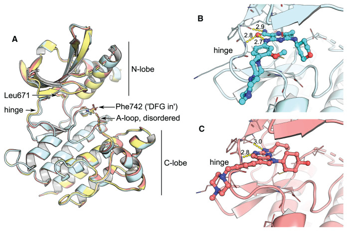
Figure 1. Comparison of unliganded MerTK with type 1 complexes.
(A) Superposition of apo-MerTK structure (PDB entry 7AB0, yellow), and complexes with gilteritinib (cyan, PDB entry 7AB1) and UNC2025 (red, PDB entry 7AB2). The compounds do not affect the overall conformation of the MerTK kinase domain or the position of the DFG motif. (B,C) Gilteritinib and UNC2025 bind to the kinase hinge without entering the back pocket. H-bonds are represented as yellow dashes with distances in Å indicated.

