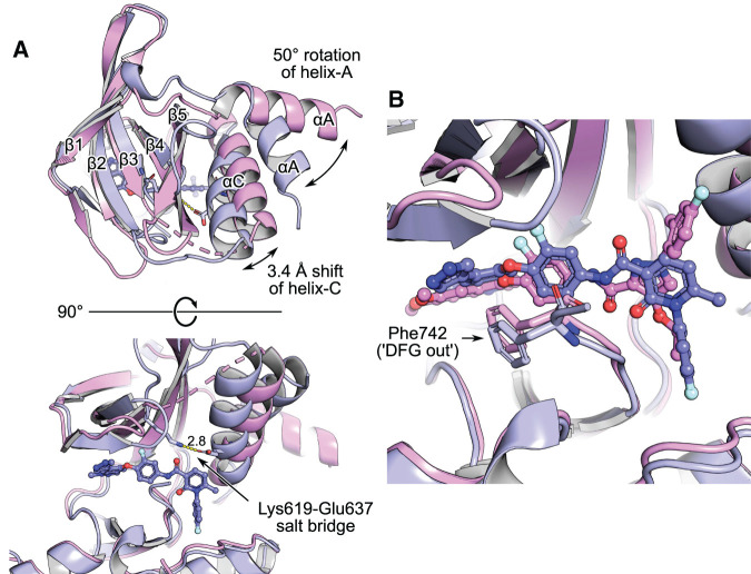
Figure 2. MerTK complexes with type 2 kinase inhibitors.
LDC1267-complex 7AAX is rendered in magenta, merestinib-complex 7AAY in purple. Alignment of the whole structures, using the CE algorithm (‘cealign’ in PyMOL). (A) Top and side view on the kinase domains. Alpha helices A and C (αA, αC) and beta strands 1–5 (β1–β5) of the kinase N-lobe are labelled. The merestinib structure depicts MerTK in ‘helix-C in’ conformation. Helix-C is repositioned 3.4 Å inward the ATP-pocket (measured on atom CA of Ser627 at the extremity of the helix), thereby enabling the formation of the canonical salt bridge between Lys619 and Glu637 (yellow dashes, distance in Å indicated). (B) Typical for type 2 compounds, both merestinib and LDC1267 displace Phe742 of the DFG motif.

