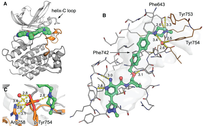
Figure 3. MerTK in complex with EX172 (PDB entry 7AAZ).
The kinase domain N-lobe is rendered in white, the C-lobe in grey and the A-loop in orange. (A) Structure overview. EX172 packs its methylpiperazine against helix-C and stabilizes the A-loop. (B) The binding pocket in detail. Residues within 5 Å of the ligand are rendered as wires and the cavity they define as surface. H-bonds are depicted as yellow dashes with distances in Å indicated. (C) Model of the structure with A-loop tyrosine 754 phosphorylated. A phosphate would replace a water molecule which is found between tyrosine hydroxyl and EX172 piperazine in the original structure 7AAZ. A phosphate would directly contact the compound, an adjacent water and Arg758.

