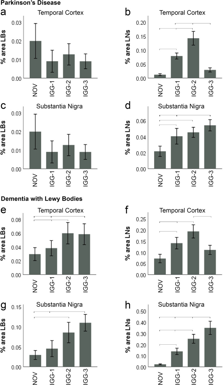
Fig. 4.
Quantification of Lewy bodies and Lewy neurites in PD and DLB. IGG-1, I GG-2, IGG-3. No difference in % area LBs in the temporal cortex and SN of PD cases was observed between each of the antibodies and NOV (a, c). b However, significantly more LNs were detected with IGG-2 and IGG-1 antibodies compared to IGG-3 and NOV (P < 0.0001); in the TC, there was no difference between IGG-3 and NOV. In addition, IGG-2 stained more LNs than IGG-1 in the temporal cortex of PD cases (P < 0.0001). d In the SN of PD cases, the % area LNs was greater for IGG-1 (P = 0.002), IGG-2 (P < 0.0001) and IGG-3 (P < 0.0001) compared to NOV. in addition IGG-3 was significantly greater than IGG-1 (P = 0.028) (d). e % area LBs detected by IGG-3 (P = 0.006) and IGG-2 (P = 0.001) was significantly greater than NOV. In addition, IGG-2 stained more LBs than IGG-1 in TC of DLB cases (P = 0.014). f Significantly more LNs were detected with IGG-2 compared to IGG-1, IGG-3 and NOV (P < 0.0001). In addition, IGG-1 stained more LNs than NOV in TC of DLB cases (P < 0.0001), but there was no difference between IGG-3 and NOV. g IGG-2 (P = 0.001) and IGG-3 (P < 0.0001) showed greater % area LBs in SN of DLB cases compared to NOV. IGG-3 also detected a higher level of LBs compared to IGG-1 (P = 0.002). h All antibodies showed significantly higher detection of LNs in SN compared to NOV in which the greatest difference occurred with IGG-3 (P < 0.0001) and the smallest with IGG-1 (P = 0.017). % area LNs with IGG-3 was also significantly higher than both IGG-1 (P < 0.0001) and IGG-2 (P = 0.001). In addition, IGG-2 was greater than IGG-1 (P = 0.013). Error bars show mean ± 95% CI

