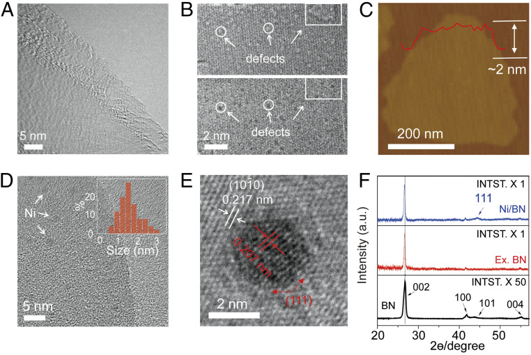
Fig. 2.
Morphology and structure characteristics of exfoliated BN nanosheets and Ni/BN nanocomposite. (A) Low-resolution transmission electron microscopy (TEM) image of typical exfoliated BN nanosheets. (B) Aberration-corrected high-resolution TEM (HRTEM) images of typical exfoliated BN nanosheet at the same location at different focus (Top, under focus; Bottom, over focus). (C) Atomic force microscopy (AFM) image of a typical exfoliated BN nanosheet. (D) The low-resolution TEM image of typical Ni nanoclusters deposited on exfoliated BN nanosheets. (Inset) Size distribution of Ni nanoclusters. (E) HRTEM images of a typical Ni nanocluster deposited on exfoliated BN nanosheets, which clearly shows the lattice structure of BN nanosheet and Ni nanocluster, respectively. (F) X-ray powder diffraction (XRD) patterns of bulk BN, exfoliated BN nanosheets, and Ni/BN nanocomposite.

