Fig. 4.
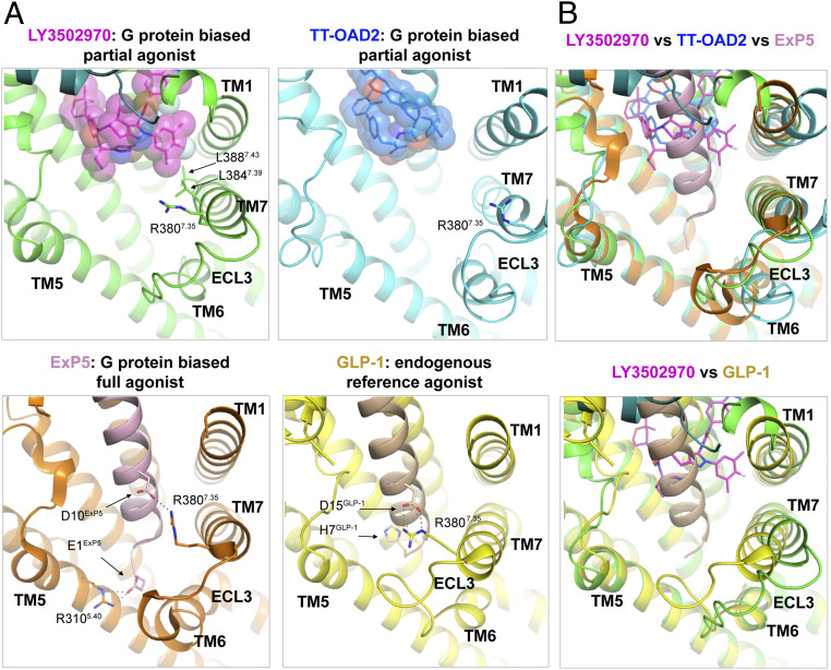
Structures of GLP-1R bound to agonists with different signaling profiles shed light on the structural basis for partial agonism and biased signaling. (A) Structures of the GLP-1R bound to LY3502970 (7TM in green, LY3502970 shown by magenta spheres), native GLP-1 (receptor in yellow, GLP-1 in beige, PDB ID code 6VCB), ExP5 (receptor in orange, ExP5 in pink, PDB ID code 6B3J), or TT-OAD2 (receptor in cyan, TT-OAD2 in blue, PDB ID code 6ORV) are aligned and shown from an identical view. Critical residues are shown by sticks. Hydrogen bonds are indicated by dashed lines. (B) The structure of GLP-1R bound to LY3502970 is aligned with GLP-1R bound to ExP5 and TT-OAD2 (Upper) and GLP-1 (Lower).

