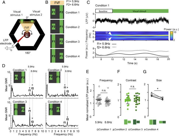
Fig. 2.
A preferred visual object evokes greater neural gain during passive viewing. (A) Open-loop paradigm. Both visual stimuli are presented to the fly in the FVF, while the LFP is recorded from the CX (Fig. 1). Arrows indicate the fly moving the ball, while visual stimuli stay fixed in the FVF. (B) Flies are presented with four flicker/position/object conditions. These conditions vary in contrast (Materials and Methods). F1 and F2 are flicker frequencies. (C, Upper) Each trial consists of a 10-s baseline and a 20-s stimulation period. (C, Lower) Example for one trial (n = 1) showing the LFP trace (top trace), the time–frequency spectrogram with increased power for both input frequencies (middle trace), and the averaged power for both input frequencies (bottom trace). (D) Signal-to-noise ratio (SNR) of power spectra averaged across all trials for all animals (f1, f2 = output frequencies of F1, F2, respectively). (E) Normalized LFP power for visual stimuli with the same input frequencies F1 = 5.9 and F2 = 6.6 for all conditions (Wilcoxon rank sum test, error bars = SEM). (F) Normalized LFP power for high- and low-contrast stimuli (Wilcoxon rank sum test, error bars = SD). (G) Normalized LFP power for large (high/low contrast) and small (high/low contrast) for all four conditions (Wilcoxon matched pair rank test). n = 6. All animals are dNPF-Gal4; UAS-CSChrimson(x)::mVenus flies that have not been fed all trans-Retinal (ATR−). n.s., not significant. *P < 0.05.

