Fig. 4.
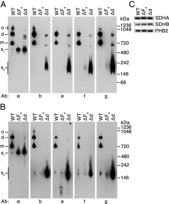
Oligomeric states of ATP synthase and vestigial complexes in HAP1 cells. Mitochondrial membranes from HAP1-WT, HAP1-ΔF6, and HAP1-Δd cells were extracted with digitonin (6 g/g protein). (A and B) Fractionation of extracts by BN-PAGE and CN-PAGE, respectively. Complexes were revealed by Western blotting with antibodies against various subunits of ATP synthase (indicated below the panels). The positions of complexes are shown on the left: o, oligomers; d, dimers; m, monomers; s1, F1-c8 subcomplex; s2, subcomplexes containing subunits b, e, f, and g. The positions of molecular weight markers are shown on the right. (C), assessment of sample loading by Western blotting of digitonin extracts, fractionated by SDS/PAGE with antibodies against the succinate dehydrogenase A (SDHA) and SDHB subunits of complex II and prohibitin 2 (PHB2).

